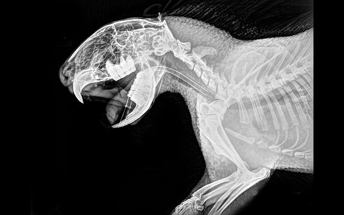
Zoo's Animal XRays Reveal Spooky, Scary Skeletons Live Science
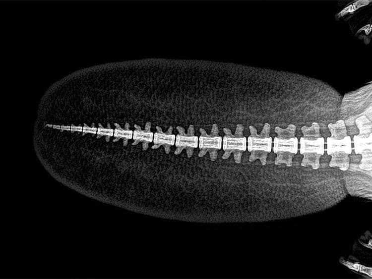
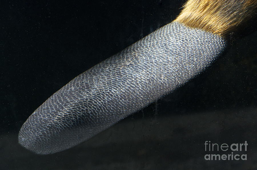
Beaver Tail, X-ray C028/1127 Rights Managed 75.4 MB (11.4 MB compressed) 6900 x 3819 pixels 58.4 x 32.3 cm · 23.0 x 12.7 in (300dpi) Request Price Add To Basket ADD TO BOARD Credit TED KINSMAN / SCIENCE PHOTO LIBRARY Caption Colour enhanced x-ray of a North American Beaver (Castor canadensis) tail.
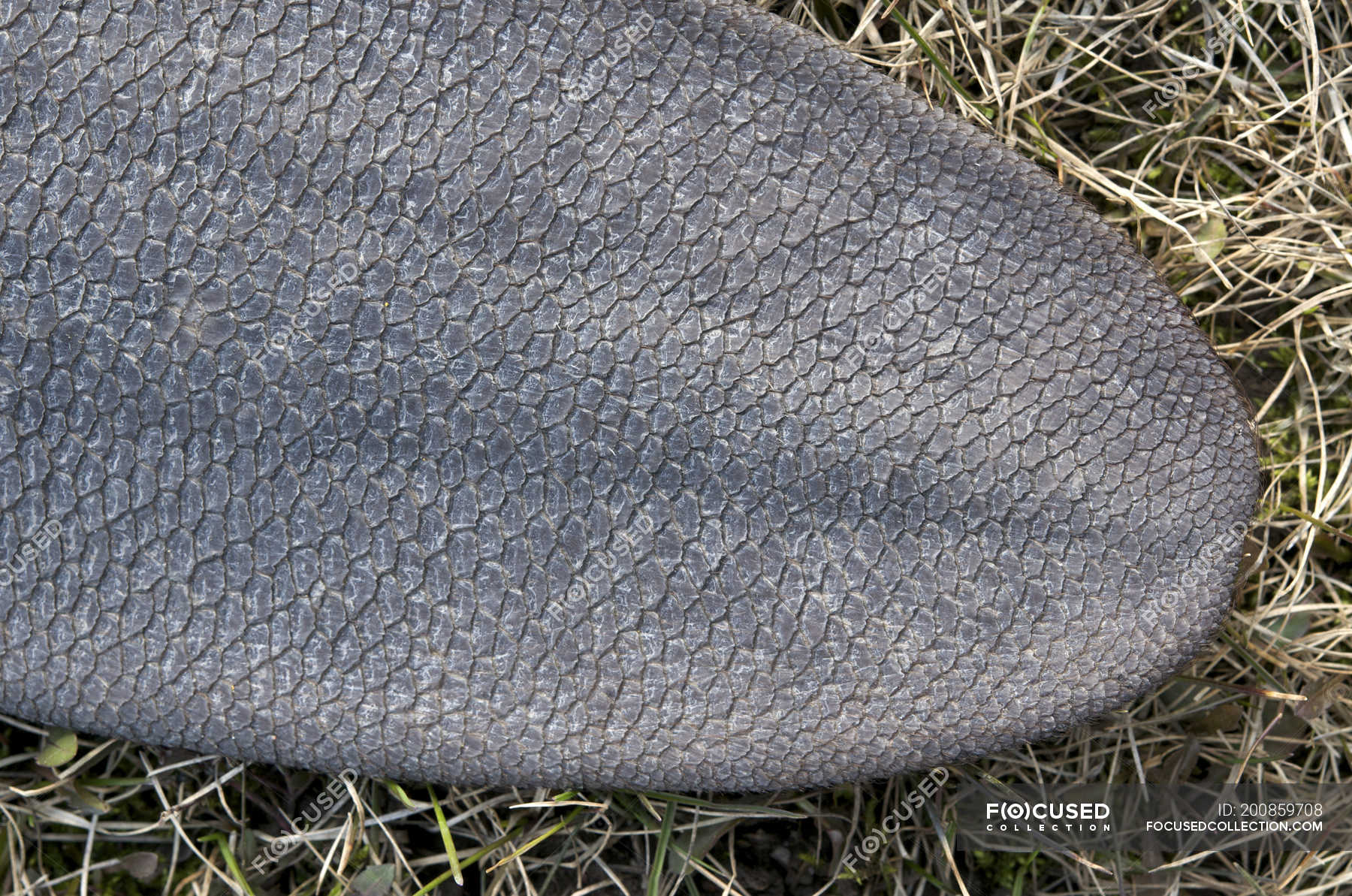
Closeup of beaver tail on dry grass — nature, selective focus Stock Photo 200859708
This anatomical variant is characterized by a prominent and elongated left lobe of the liver, resembling the shape of a beaver's tail. It is considered a benign and asymptomatic condition, discovered incidentally during imaging studies.
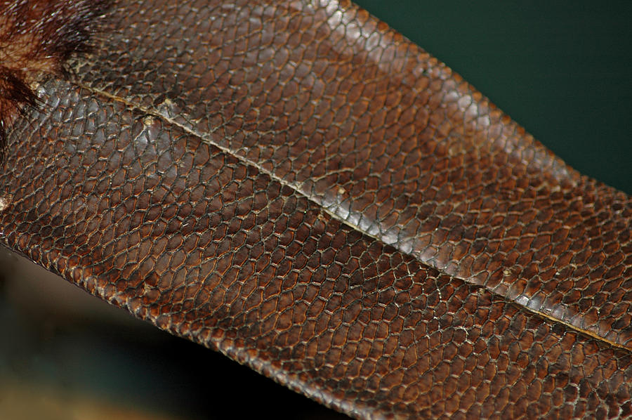
Beaver Tail Photograph by LeeAnn McLaneGoetz
This is a fibrous band which attaches to the diaphragm at the left extremity of the liver and may contain liver parenchyma. 2 This is an important structure intraoperatively as it may contain blood vessels and bile ducts and should be ligated with care. 1 In a cadaveric study, for example, 'comparatively large bile ducts' were found in 12 of 27.
Examples of American beaver tails when transmitters pulled back through... Download Scientific
Beaver tail liver is an anatomical liver variant, that can be misdiagnosed as a perisplenic hemorrhage or a subcapsular hematoma within the splenic parenchyma. Reviewing the anatomy in multiple planes aids in making the correct diagnosis, as does reviewing other modalities such as ultrasound and MRI. 1 article features images from this case

Beaver tail liver
Beaver tail liver is a rare hepatic anatomical variant in which the left hepatic lobe extends into the left upper quadrant and surrounds the spleen. This extension of the left hepatic lobe consists of normal hepatic parenchyma with no functional liver impairment.

Figure 1 from Beaver tail liver on pediatric chest Xray Semantic Scholar
Beaver tail liver, or else known as the sliver of liver, is a rare anatomic variation of the liver where the left lobe of the liver extends laterally to contact and enwrap the spleen. A case is presented here where a middle-aged male presented with complaints of abdominal pain, hematuria, and fever.
15 Thrilling Animal XRays from the Oregon Zoo
Europe PMC is an archive of life sciences journal literature.

Closeup of beaver tail. (Castor canadensis). Northern Ontario, Canada Stock Photo Alamy
Age: 25 years Gender: Male X-Ray Chest P/A View x-ray Frontal Frontal Small area of pulmonary opacity near left costophrenic angle. Fundic shadow of stomach displaced inferiorly and medially by what appears like a thick homogeneous opacity having a curved lateral end.
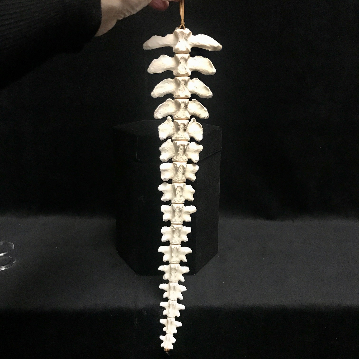
Fascinating, articulated beaver tail bones, available at natur
Beaver tail liver is a rare hepatic anatomical variant in which the left hepatic lobe extends into the left upper quadrant and surrounds the spleen. This extension of the left hepatic lobe consists of normal hepatic parenchyma with no functional liver impairment. In trauma cases, however, the extended left hepatic lobe is vulnerable to injury.
(PDF) A rare case of left diaphragmatic palsy with beaver tail anomaly of liver mimicking
Beaver tail liver, also known as a sliver of liver, is a variant of normal hepatic morphology with an elongated left liver lobe extending laterally up to the left hypochondrium and often surrounding the spleen. [ 1] The term was coined because of its resemblance to a beaver's tail.
My cat has had a broken tail since birth, finally got an xray done on it a few years later
Beaver tail liver is an anatomical liver variant presenting as elongated left lobe of liver which extends laterally to the spleen. It can present with symptoms or be detected accidentally.

All Categories Laurens Wildlife Rescue
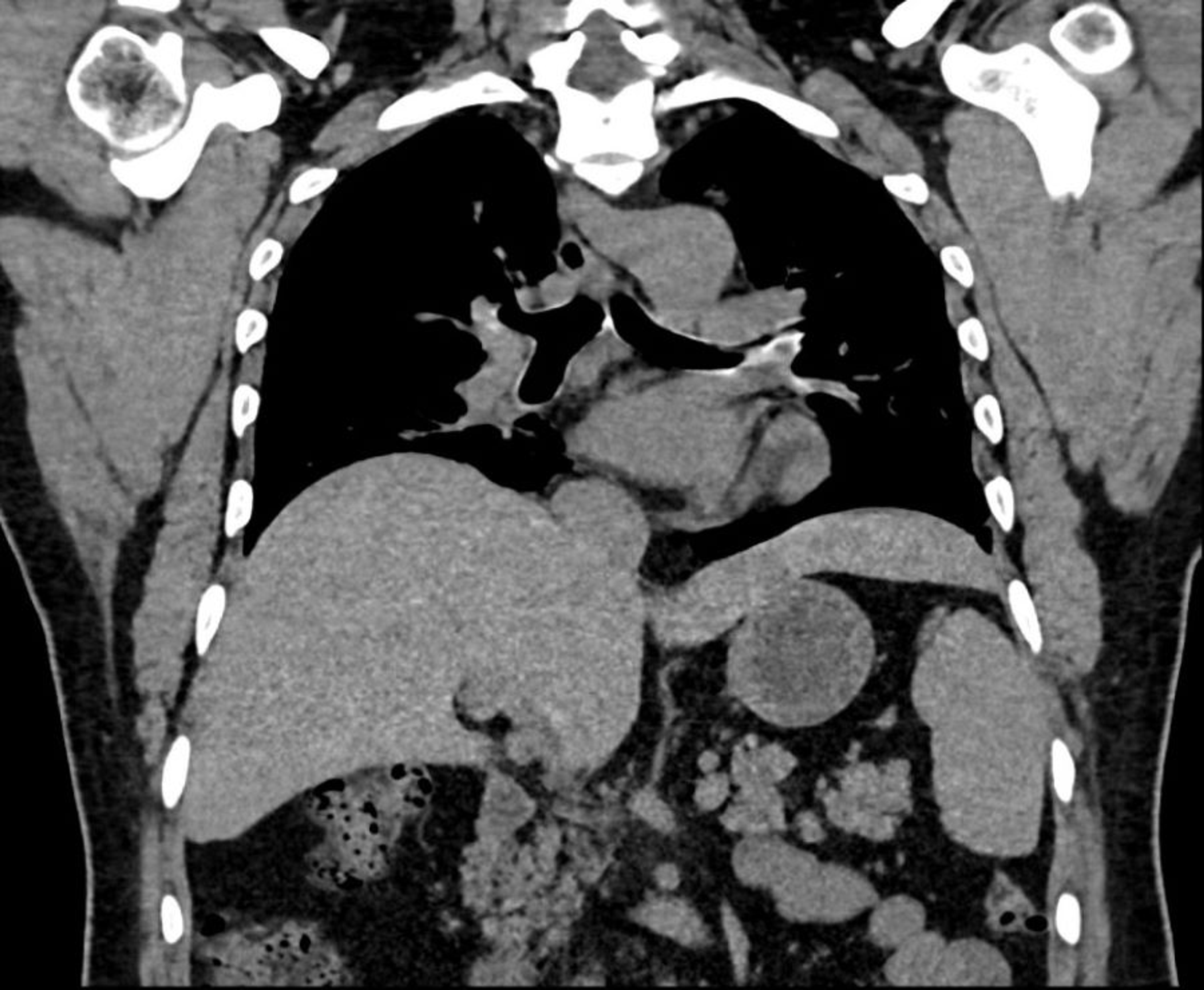
Patient Data Age: 35 years Gender: Female ct Coronal and axial CT images show the inferior lobe of the liver lying below the costal margin, and elongated left liver lobe extends laterally and in close contact with the spleen. Case Discussion

Beaver Tail Stock Photo Image 49145217
Gross anatomy. The liver is an irregular, wedge-shaped organ that lies below the diaphragm in the right upper quadrant of the abdominal cavity and is in close approximation with the diaphragm, stomach and gallbladder.It is largely covered by the costal cartilages 9.. The liver is made of several functional units called lobules, which in turn can be subdivided into smaller units called sinusoids.

This Is What Animal XRays Look Like
Beaver tail liver is an anatomical liver variant presenting as elongated left lobe of liver which extends laterally to the spleen. It can present with symptoms or be detected accidentally.
Cureus Beaver Tail Liver A Hepatic Morphology Variant
RadiologyCaseReports17(2022)4780-4783 4781 Fig. 1 -The image is showing chest X-ray done at first patient evaluation, with the red arrow pointing to the shadow left
Beaver Tail Photograph by Sean Griffin Fine Art America
Liver / diagnostic imaging* Tomography, X-Ray Computed / methods* Ultrasonography / methods*